CT Colonography
Computed tomography (CT) colonography or virtual colonoscopy uses special x-ray equipment to examine the large intestine for cancer and growths called polyps. During the exam, the doctor inserts a small tube a short distance into the rectum. The doctor uses gas or air to inflate the colon and the rectum and takes pictures.
Prior to your exam, your doctor may restrict you to clear fluids on the day before the CT and give you instructions on clearing your bowels. Tell your doctor if there's a possibility you are pregnant and discuss any recent illnesses, medical conditions, medications you're taking, and allergies. Your doctor will tell you not to eat or drink anything for a few hours beforehand. If you have a known allergy to contrast material, your doctor may prescribe medications to reduce the risk of an allergic reaction. You must take these medications 12 hours prior to your exam. Leave jewelry at home and wear loose, comfortable clothing. You will need to wear a gown.
- What is CT Colonography?
- What are some common uses of the procedure?
- How should I prepare?
- What does the equipment look like?
- How does the procedure work?
- How is the procedure performed?
- What will I experience during and after the procedure?
- Who interprets the results and how do I get them?
- What are the benefits vs. risks?
- What are the limitations of CT Colonography?
What is CT Colonography?
Computed tomography, more commonly known as a CT or CAT scan, is a diagnostic medical imaging test. Like traditional x-rays, it produces multiple images or pictures of the inside of the body.
A CT scan generates images that can be reformatted in multiple planes. It can even generate three-dimensional images. Your doctor can review these images on a computer monitor, print them on film or via a 3D printer, or transfer them to a CD or DVD.
CT images of internal organs, bones, soft tissue, and blood vessels provide greater detail than traditional x-rays. This is especially true for soft tissues and blood vessels.
CT colonography, also known as virtual colonoscopy, uses low dose radiation CT scanning to obtain an interior view of the colon (the large intestine). This area is otherwise only seen with a more invasive procedure where the doctor inserts an endoscope into the rectum and passes it through the entire colon.
What are some common uses of the procedure?
The major reason for performing CT colonography is to screen for polyps or cancers in the large intestine. Polyps are growths that arise from the inner lining of the intestine. A very small number of polyps may grow and turn into cancers.
The goal of screening with CT colonography is to find these growths in their early stages. This allows the doctor to remove them before cancer has a chance to develop. The American Cancer Society (ACS) recommends that women and men undergo screening for colon cancer or polyps beginning at age 45. As part of its recommendation, ACS suggests CT colonography as an option once every five years. Individuals at increased risk or with a family history of colon cancer may start screening at age 40 or younger and may be screened at shorter intervals (for example, having a colonoscopy every five years). Risk factors for the disease include a history of polyps or having a family history of colon cancer. Signs and symptoms of colon cancer include a persistent change in bowel habits, the presence of blood in the stool, abdominal discomfort or pain, bloating and unexplained weight loss.
How should I prepare?
You should wear comfortable, loose-fitting clothing to your exam. You will need to wear a gown during the procedure.
Women should always inform their physician and the CT technologist if there is any possibility that they may be pregnant. See the CT Safety During Pregnancy page for more information.
The bowel-cleansing regimen for CT colonography may be similar to that for a colonoscopy or consist of a smaller volume of cleansing liquid. Your diet will be restricted to clear liquids the day before the exam. It is very important to clean out your colon the night before your exam so that the radiologist can clearly see any polyps that might be present. You will take either a set of pills and/or a liquid laxative. Some common preparations are Magnesium Citrate and bisacodyl tablets and/or NuLytely®, Go-Lytely® (Polyethylene glycol electrolyte solutions). You may take additional agents the day before the exam. These may include small quantities of barium and iodinated liquids. These agents help the radiologist better distinguish stool from polyps by "tagging" the remaining stool and fluid.
Be sure to tell your doctor if you have heart, liver, or kidney disease to be certain that the bowel prep will be safe. Your doctor can advise you on dietary restrictions prior to the exam. You will be able to resume your usual diet immediately after the exam.
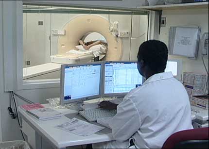
What does the equipment look like?
The CT scanner is typically a large, donut-shaped machine with a short tunnel in the center. You will lie on a narrow table that slides in and out of this short tunnel. Rotating around you, the x-ray tube and electronic x-ray detectors are located opposite each other in a ring, called a gantry. The computer workstation that processes the imaging information is in a separate control room. This is where the technologist operates the scanner and monitors your exam in direct visual contact. The technologist will be able to hear and talk to you using a speaker and microphone.
During CT colonography, you will lie on your back and then on your stomach and/or side. Sometimes, the doctor may ask you to lie on just the right and left sides. Before starting the exam, be sure to let the staff know if you have any limitations in your ability to move.
How does the procedure work?
In many ways, a CT scan works like other x-ray exams. X-rays are a form of radiation–like light or radio waves–that can be directed at the body. Different body parts absorb x-rays in different amounts. This difference allows the doctor to distinguish body parts from one another on an x-ray or CT image.
In a conventional x-ray exam, the technologist directs a small amount of radiation through the part of the body under examination. A special electronic image recording plate captures the image. Bones appear white on the x-ray. Soft tissue, such as the heart or liver, shows up in shades of gray. Air appears black.
With CT scanning, several x-ray beams and electronic x-ray detectors rotate around you. These measure the amount of radiation being absorbed throughout your body. Sometimes, the exam table will move during the scan, so that the x-ray beam follows a spiral path. A special computer program processes this large volume of data to create two-dimensional cross-sectional images of your body. These images are displayed on a monitor. CT imaging is sometimes compared to looking into a loaf of bread by cutting the loaf into thin slices. When the image slices are reassembled by computer software, the result is a very detailed multidimensional view of the body's interior.
Refinements in detector technology allow nearly all CT scanners to obtain multiple slices in a single rotation. These multi-slice or multidetector CT scanners obtain thinner slices in less time. This results in more detail and additional view capabilities.
Modern CT scanners can scan through large sections of the body in just a few seconds. Such speed is beneficial for all patients but especially children, the elderly, and critically ill.
For CT colonography, the computer generates a detailed three-dimensional (3-D) model of the colon, which the radiologist uses to view the bowel in a way that simulates traveling through the colon. This is why the procedure is often called a virtual colonoscopy. Two-dimensional (2-D) images of the inside of the colon as well as the rest of the abdomen and pelvis are obtained and reviewed at the same time without any additional radiation.
How is the procedure performed?
The technologist begins by positioning you on the CT exam table, usually lying flat on your back. They may use straps and pillows to help you maintain the correct position and remain still during the exam.
The doctor will pass a very small, flexible tube two inches into your rectum and gently pump air into the colon using a hand-held squeeze bulb. Sometimes an electronic pump delivers carbon dioxide gas into the colon. Sometimes the doctor inflates a small balloon on the rectal tube to help keep the tube positioned correctly. The purpose of the gas is to distend (inflate) the colon as much as possible to eliminate any folds or wrinkles that might hide polyps from the doctor’s view.
Next, the table will move through the scanner. The technologist may ask you to hold your breath for about 15 seconds before turning over and lying on your back or side for a second pass through the scanner. In some centers, the sequence of positions may be the opposite: facing upward first and then facing down. Once the scan is done, the doctor or technologist will remove the tube.
The entire exam usually takes about 15 minutes.
What will I experience during and after the procedure?
Most patients who have CT colonography report a feeling of fullness when the colon is inflated during the exam, as if they need to pass gas. Significant pain is uncommon, occurring in fewer than 5 percent of patients. The doctor or technologist may inject a muscle-relaxing drug intravenously or subcutaneously (under the skin) to lessen discomfort, but this is seldom necessary. The scanning procedure itself causes no pain or other symptoms.
When you enter the CT scanner, you may see special light lines projected onto your body. These lines help ensure that you are in the correct position on the exam table. With modern CT scanners, you may hear slight buzzing, clicking and whirring sounds. These occur as the CT scanner's internal parts, not usually visible to you, revolve around you during the imaging process.
You will be alone in the exam room during the CT scan, unless there are special circumstances. For example, sometimes a parent wearing a lead shield may stay in the room with their child. However, the technologist will always be able to see, hear and speak with you through a built-in intercom system.
After a CT exam, you can return to your normal activities.
Who interprets the results and how do I get them?
A radiologist, a doctor specially trained to supervise and interpret radiology exams, will analyze the images. The radiologist will send an official report to the doctor who ordered the exam.
In some cases, information about whether you have polyps is available immediately. Some imaging centers can perform colonoscopy and, if necessary, polyp removal the same day as the CT colonography.
You may need a follow-up exam. If so, your doctor will explain why. Sometimes a follow-up exam further evaluates a potential issue with more views or a special imaging technique. It may also see if there has been any change in an issue over time. Follow-up exams are often the best way to see if treatment is working or if a problem needs attention.
What are the benefits vs. risks?
Benefits
- This minimally invasive test provides both 2-D and 3-D images that can depict many polyps and other lesions as clearly as when they are directly seen by conventional colonoscopy.
- CT colonography has a much lower risk of perforating the colon than conventional colonoscopy. Most people who undergo CT colonography do not have polyps and can be spared having to undergo a full colonoscopy which typically requires sedation.
- CT colonography is an excellent alternative for patients who have clinical factors that increase the risk of complications from colonoscopy, such as treatment with a blood thinner or a severe breathing problem.
- Elderly patients, especially those who are frail or ill, will tolerate CT colonography better than conventional colonoscopy.
- CT colonography can be helpful when colonoscopy cannot be completed because the bowel is narrowed or obstructed for any reason, such as by a large tumor.
- If conventional colonoscopy cannot reach the full length of the colon—which occurs up to 10 percent of the time—CT colonography can be performed on the same day because the colon has already been cleansed.
- CT colonography provides clearer and more detailed images than a conventional barium enema x-ray examination.
- CT colonography can detect abnormalities outside of the colon, including early-stage malignancies in other organs and potentially dangerous conditions, such as abdominal aortic aneurysms.
- CT colonography is well tolerated. Sedation and pain relievers are not needed, so there is no recovery period and you can return to your normal daily activities immediately after the test.
- CT colonography is less costly than colonoscopy.
- No radiation remains in a patient's body after a CT exam.
- The x-rays used for CT scanning should have no immediate side effects.
Risks
- There is a very small risk that inflating the colon with air could injure or perforate the bowel. This has been estimated to happen in fewer than one in 10,000 patients.
- There is always a slight chance of cancer from excessive exposure to radiation. However, the benefit of an accurate diagnosis far outweighs the risk involved with CT scanning.
- The radiation dose for this procedure varies. See the Radiation Dose page for more information.
- Women should always tell their doctor and x-ray or CT technologist if there is any chance they are pregnant. See the Radiation Safety page for more information about pregnancy and x-rays.
- Doctors do not generally recommend CT scanning for pregnant women unless medically necessary because of potential risk to the unborn baby.
What are the limitations of CT Colonography?
A person who is very large may not fit into the opening of a conventional CT scanner. Or, they may be over the weight limit—usually 450 pounds—for the moving table.
CT colonography is strictly a diagnostic procedure. If any clinically significant polyps are found, your doctor will have to remove them using conventional colonoscopy.
CT colonography may not differentiate stool from artifacts and smaller polyps as well as conventional colonoscopy.
CT colonography is not recommended for patients who have active Crohn's disease, ulcerative colitis, inflammatory bowel disease or diverticulitis, because of increased risk of perforating the colon. Patients with a history of bowel perforation and those experiencing severe pain or cramps on the day of the exam should not undergo CT colonography.
Some insurance companies do not cover CT colonography as a screening test for colonic polyps, but they may cover the cost if a patient has symptoms related to the colon. Recently, the United States Preventive Services Task Force (USPSTF) assigned a grade of "A" to CT colonography as a screening modality for the detection of colon cancer. This decision, according to provisions included in the Affordable Care Act, mandates coverage by private insurance companies participating in the insurance exchange program. Medicare does not currently cover CT colonography for screening but may do so in the near future due to the decision of the USPSTF.
Additional Information and Resources
RTAnswers.org: Radiation Therapy for Colorectal Cancer
This page was reviewed on November 01, 2022