Stereotactic Breast Biopsy
Stereotactic breast biopsy uses mammography – a specific type of breast imaging that uses low-dose x-rays — to help locate a breast abnormality and remove a tissue sample for examination under a microscope. It's less invasive than surgical biopsy, leaves little to no scarring and can be an excellent way to evaluate calcium deposits or tiny masses that are not visible on ultrasound.
Tell your doctor if there's a possibility you are pregnant. Discuss any medications you're taking, including aspirin and herbal supplements, and whether you have any allergies — especially to anesthesia. You will be advised to stop taking aspirin, blood thinners, or particular herbal supplements which can increase your risk of bleeding for three to five days before your procedure. Don't wear deodorant, talcum powder or lotion under your arms or on your breasts for your procedure as these may appear on the mammogram. Leave jewelry at home and wear loose, comfortable clothing. You may be asked to wear a gown.
- What is Stereotactic (Mammographically Guided) Breast Biopsy?
- What are some common uses of the procedure?
- How should I prepare?
- What does the equipment look like?
- How does the procedure work?
- How is the procedure performed?
- What will I experience during and after the procedure?
- Who interprets the results and how do I get them?
- What are the benefits vs. risks?
- What are the limitations of Stereotactic Breast Biopsy?
What is Stereotactic (Mammographically Guided) Breast Biopsy?
Physical, mammography, and other exams often detect lumps or abnormalities in the breast. However, these tests cannot always tell whether a growth is benign or cancerous.
Doctors use breast biopsy to remove a small amount of tissue from a suspicious area for lab analysis. The doctor may perform a biopsy surgically. More commonly, a radiologist will use a less invasive procedure that involves a hollow needle and image-guidance. Image-guided needle biopsy does not remove the entire lesion. Instead, it obtains a small sample of the abnormality for further analysis.
Image-guided biopsy uses ultrasound, MRI, or mammography imaging guidance to take samples of an abnormality.
In stereotactic breast biopsy, a special mammography machine uses x-rays to help guide the radiologist's biopsy equipment to the site of the imaging abnormality.
What are some common uses of the procedure?
A stereotactic breast biopsy may be performed when a mammogram shows a breast abnormality such as:
- a suspicious mass
- tiny clusters of small calcium deposits (microcalcifications)
- a distortion in the structure of the breast tissue
- an area of abnormal tissue change
- a new mass or area of calcium deposits in a previous surgery site.
Stereotactic breast biopsy is performed as a non-surgical method of assessing a breast abnormality. If the results show cancer cells, the surgeon can use this information for planning treatment.
How should I prepare?
You may need to remove some clothing and/or change into a gown for the exam. Remove jewelry, removable dental appliances, eyeglasses, and any metal objects or clothing that might interfere with the x-ray images.
Women should always tell their doctor if there is any possibility that they are pregnant. Doctors do not perform some procedures using imaging guidance during pregnancy because radiation can be harmful to the fetus.
You should not wear deodorant, powder, lotion or perfume under your arms or on your breasts on the day of the exam.
Prior to a needle biopsy, you should report to your doctor all medications that you are taking, including herbal supplements, and if you have any allergies, especially to anesthesia. Your physician may advise you to stop taking aspirin, blood thinners, or certain herbal supplements for three to five days before your procedure to decrease your risk of bleeding. Also, inform your doctor about recent illnesses or other medical conditions.
What does the equipment look like?
The specialized mammography machine used in this procedure is similar to the mammography unit used to produce mammograms.
A mammography unit is a box with a tube that produces x-rays. The unit is used exclusively for breast x-ray exams and features special accessories to limit x-ray exposure to only the breast. The unit features a device to hold and compress the breast and position it so the technologist can capture images at different angles.
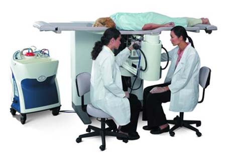
At most facilities, a specially designed examination table will allow you to lie face down with your breast hanging freely through an opening in the table. The table is then raised and the biopsy procedure is performed beneath the table. At other facilities, the procedure may be performed while you sit in a chair.
Tissue sample is obtained using:
- A vacuum-assisted device (VAD), a vacuum powered instrument that uses pressure to pull tissue into the needle. This instrument rotates positions and collects multiple tissue samples through one needle insertion.
Other sterile equipment involved in this procedure includes syringes, sponges, forceps, scalpels and a specimen cup or microscope slide.
How does the procedure work?
Mammography is a low-dose x-ray system designed to evaluate breast tissue.
A special digital mammography machine is used to perform a stereotactic breast biopsy. In digital mammography, as in digital photography, film is replaced by electronic detectors. These convert x-rays into electrical signals, which are used to produce images of the breast that can be immediately seen on a computer screen.
Stereotactic mammography pinpoints the exact location of a breast abnormality by using computer analysis of x-rays taken from two different angles. Using the calculated computer coordinates, the radiologist inserts the needle through a small cut in the skin, then advances it into the lesion and removes tissue samples.
How is the procedure performed?
Image-guided, minimally invasive procedures such as stereotactic breast biopsy are most often performed by a specially trained radiologist.
Breast biopsies are usually done on an outpatient basis.
In most cases, you will lie face down on a moveable exam table. The doctor will position the affected breast into an opening in the table.
The table is raised and the procedure is then performed beneath it. If the machine is an upright system, you may be seated in front of the stereotactic mammography unit.
The breast is compressed and held in position throughout the procedure.
Preliminary stereotactic mammogram images are taken and reviewed by the radiologist. Once the radiologist identifies the abnormality on imaging, the computer will generate coordinate information and send it to the biopsy device.
The doctor will inject a local anesthetic into the skin and more deeply into the breast to numb it.
The doctor will make a very small nick in the skin at the site where they will insert the biopsy needle.
The radiologist then inserts the needle and advances it to the location of the abnormality using the mammogram and computer generated coordinates. Mammogram images are again obtained to confirm that the needle is within the lesion prior to sampling.
Tissue samples are then removed, generally using a vacuum-assisted device. Typically, three to twelve samples are obtained, depending on the device used.
If calcium deposits (calcifications) are being sampled, an x-ray of the removed tissue will be obtained to document enough deposits were obtained for analysis under a microscope. Additional sampling may be needed if not enough calcifications are identified initially.
After the sampling is complete, the needle will be removed from the breast.
A final set of images will be taken.
The doctor may place a small marker at the biopsy site so they can locate it in the future if necessary.
Once the biopsy is complete, the doctor or nurse will apply pressure to stop any bleeding. They will cover the opening in the skin with a dressing. No sutures are needed.
The doctor may use mammography to confirm that the marker is in the proper position.
This procedure is usually completed within an hour.
What will I experience during and after the procedure?
You will be awake during your biopsy and should have little discomfort. Many women report little pain and no scarring on the breast. However, certain patients, including those with dense breast tissue or abnormalities near the chest wall or behind the nipple, may be more sensitive during the procedure.
Some women find that the major discomfort of the procedure is from lying on their stomach for the length of the procedure. Strategically placed cushions can ease this discomfort. Some women may also experience neck and/or back pain as the head is turned to the side when the doctor positions the breast for biopsy.
When you receive the local anesthetic to numb the skin, you will feel a pin prick from the needle followed by a mild stinging sensation from the local anesthetic. You will likely feel some pressure when the doctor inserts the biopsy needle and during tissue sampling. This is normal.
The area will become numb within a few seconds.
You must remain very still while the doctor performs the imaging and the biopsy.
As tissue samples are taken, you may hear clicks or buzzing sounds from the sampling instrument. These are normal.
If you experience swelling and bruising following your biopsy, your doctor may tell you to take an over-the-counter pain reliever and to use a cold pack. Temporary bruising is normal.
Call your doctor if you experience excessive swelling, bleeding, drainage, redness, or heat in the breast.
If a marker is left inside the breast to mark the location of the biopsied lesion, it will cause no pain, disfigurement, or harm. Biopsy markers are MRI compatible and will not cause metal detectors to alarm.
Avoid strenuous activity for at least 24 hours after the biopsy. Your doctor will outline more detailed post-procedure care instructions for you.
Who interprets the results and how do I get them?
A pathologist examines the removed specimen and makes a final diagnosis. Depending on the facility, the radiologist or your referring physician will share the results with you. The radiologist will also evaluate the results of the biopsy to make sure that the pathology and image findings explain one another. In some instances, even if cancer is not diagnosed, surgical removal of the entire biopsy site and imaging abnormality may be recommended if the pathology does not match the imaging findings.
You may need a follow-up exam. If so, your doctor will explain why. Sometimes a follow-up exam further evaluates a potential issue with more views or a special imaging technique. It may also see if there has been any change in an issue over time. Follow-up exams are often the best way to see if treatment is working or if a problem needs attention.
What are the benefits vs. risks?
Benefits
- The procedure is less invasive than surgical biopsy, leaves little or no scarring, and can be performed in less than an hour.
- Stereotactic breast biopsy is an excellent way to evaluate calcium deposits or masses that are not visible on ultrasound.
- Stereotactic core needle biopsy is a simple procedure that may be performed in an outpatient imaging center.
- Compared with open surgical biopsy, the procedure is about one-third the cost.
- Very little recovery time is required.
- Generally, the procedure is not very painful.
- No breast defect remains and, unlike surgery, stereotactic needle biopsy does not distort the breast tissue or make it difficult to read future mammograms.
- Recovery time is brief and patients can soon resume their usual activities.
- No radiation stays in your body after an x-ray exam.
- X-rays usually have no side effects in the typical diagnostic range for this exam.
Risks
- There is a risk of bleeding and forming a hematoma, or a collection of blood at the biopsy site. The risk, however, appears to be less than one percent of patients.
- An occasional patient has significant discomfort, which can be readily controlled by non-prescription pain medication.
- Any procedure where the skin is penetrated carries a risk of infection. The chance of infection requiring antibiotic treatment appears to be less than one in 1,000.
- Depending on the type of biopsy or the design of the biopsy machine, a biopsy of tissue located deep within the breast carries a slight risk that the needle will pass through the chest wall. This could allow air around the lung and cause the lung to collapse. This is extremely rare.
- There is a small chance that this procedure will not provide the final answer to explain the imaging abnormality.
- There is always a slight chance of cancer from excessive exposure to radiation. However, given the small amount of radiation used in medical imaging, the benefit of an accurate diagnosis far outweighs the associated risk.
- Women should always tell their doctor and x-ray technologist if they are pregnant. See the Radiation Safety page for more information about pregnancy and x-rays.
What are the limitations of Stereotactic Breast Biopsy?
There are some instances in which stereotactic biopsy may not be possible, including if:
- The target abnormality is located near the chest wall or directly behind the nipple.
- The mammogram shows only a vague change in tissue density but no definite mass or nodule. The finding may be too subtle to identify at time of biopsy.
- The breast is too thin.
- The target is composed of diffuse calcium deposits scattered throughout the breast, which on occasion are difficult to target.
Breast biopsy procedures will occasionally miss a lesion or underestimate the extent of disease present. If the diagnosis remains uncertain after a technically successful procedure, surgical biopsy will usually be necessary.
Additional Information and Resources
RTAnswers.org: Radiation Therapy for Breast Cancer
Society of Interventional Radiology (SIR) - Patient Center
This page was reviewed on March 11, 2024


